


What is the procedure for removing a mole from the skin?
The procedure for removing a mole from the skin involves surgically or ablative (burning) removal of skin lesions that appear different from the normal skin color and are sometimes raised brown or other colored spots on the skin.
How is mole removal Turkey done?
The most commonly used technique for mole removal is the excision + primary closure technique. With this technique, the surgeon makes an incision around the mole to be removed, under local (regional) or general anesthesia. The incision is made to encompass the base of the mole as well. Afterward, the bleeding is stopped, and the wound is closed with stitches. The benefit of this method is that the removed tissue can be sent for pathological examination to determine whether the mole is benign or malignant.
Alternatively, ablative methods can also be used in mole removal Turkey. In this method, the mole is directly burned using devices such as radiofrequency (RF), plasma pen, or CO2 laser. After being burned, the upper layers of the skin are removed, and if necessary, the deeper layers are burned again. This leaves a burned area resembling a crater, which will heal on its own. Antibiotics and reparative creams are applied during the healing process. This method is particularly preferred for small and raised lesions on the skin as it eliminates the risks associated with sutures, suture scars, and suture dehiscence. However, the mole that has been burned cannot be sent for pathological examination, and skin cancer may be overlooked.
How long does a mole removal Turkey surgery take?
The duration of a mole removal Turkey procedure can vary. It can take only seconds for polypoid moles like “skin tags,” while it can take up to 30 minutes for subcutaneous moles with sebaceous gland components. On average, the removal of a mole takes about 5-10 minutes.
Is mole removal Turkey a painful procedure?
The area is numbed with local or general anesthesia, so there is no pain. However, the patient may feel some pulling sensations if they are awake during the procedure. But it is not painful. After the procedure, painkillers are prescribed. If these painkillers are taken before the numbness wears off, there shouldn’t be significant pain after the procedure. It may feel like a slight stinging sensation, similar to when we get a paper cut on our fingers.
How is the healing process after mole removal?
Using ablative methods depends on the size of the wound and factors related to the patient (age, overall health, smoking habits, vascularity of the operated area). For example, in a young and healthy individual with a 3-4mm mole on the face that is burned or ablated, healing is expected within 7-10 days. However, for an 80-year-old individual with a 3-4cm mole on the foot, suffering from heart failure and asthma, the wound healing may take months and require additional treatments.
Under normal circumstances, a mole wound on the face that is excised and sutured will result in the skin healing within 5-10 days. Generally, facial stitches are removed within 5-7 days, and scar-reducing creams are recommended (such as centella extract, silicone gel, SPF 50+ sunscreen, etc.). However, this process can vary depending on the individual’s overall health, age, underlying conditions, location of the procedure, smoking habits, wound tension, and other factors. Wounds that are tightly sutured, wounds in highly mobile areas (such as the area around the mouth), wounds that remain moist (such as the armpit area due to sweating), infected wounds, and wounds of diabetic patients can all lead to suture dehiscence and prolong the healing process. Individuals with high blood pressure may experience oozing bleeding in the first few days after the procedure, and blood may accumulate within the wound. While applying pressure and cold compress is usually sufficient to stop it, persistent bleeding may require additional interventions from the surgeon.
On average, after a mole removal Turkey procedure, there may be swelling and bruising for the first 2 days, and the stitches are typically removed within 7-10 days. Bruising will fade during this time. Within 1-2 months, the scar will transition from a dark color to pink and become less noticeable. If there are subcutaneous stitches, the body may extrude them within the first 6 months (a phenomenon known as stitch spitting). If there is tension on the skin or if ablative methods were used, the scar may widen or narrow, respectively, within the first year. Between 6-18 months, the scar will match the surrounding skin tone. It’s important to protect the area from the sun during this process.
You can return to work 2 days after the procedure. However, if you have a physically demanding job, we recommend returning to work after 1 week. Regardless of your occupation, we advise protecting the surgical area from trauma for 6 months.
Does mole removal leave scars? How can scars be minimized?
Any procedure involving a full-thickness incision in the skin can leave some degree of scarring. Especially in areas with low tension and thin skin, such as around the eyes, the scar may be less noticeable. However, in areas with thick and tight skin, such as the shoulders or the area between the breasts, the scars can be more pronounced. Plastic surgeons aim to minimize scars by using techniques that reduce tension on the wound, such as using superficial or subcutaneous sutures depending on the location of the incision. They may choose sutures that will dissolve or need to be removed at a later time. For small lesions, the procedure can be done using a shave excision method, which involves superficially shaving off the lesion without suturing. Alternatively, if a full-thickness incision is made, the surgeon can use layered sutures, placing separate sutures in the fascia, subcutaneous tissue, and skin, to prevent the incision from widening during the healing process.
What are the risks and possible complications of the removal procedure?
A person who undergoes mole removal Turkey (nevus excision) must understand and accept the risks of general surgery. When a part of the body is cut and stitched, complications such as bleeding, infection, wound healing problems, scarring, and pain due to the procedure can be seen in this area. These risks are the risks that can be seen in all surgical procedures. Before performing the procedure, a consent form is filled in that you accept the risks and complications of the mole removal Turkey procedure.
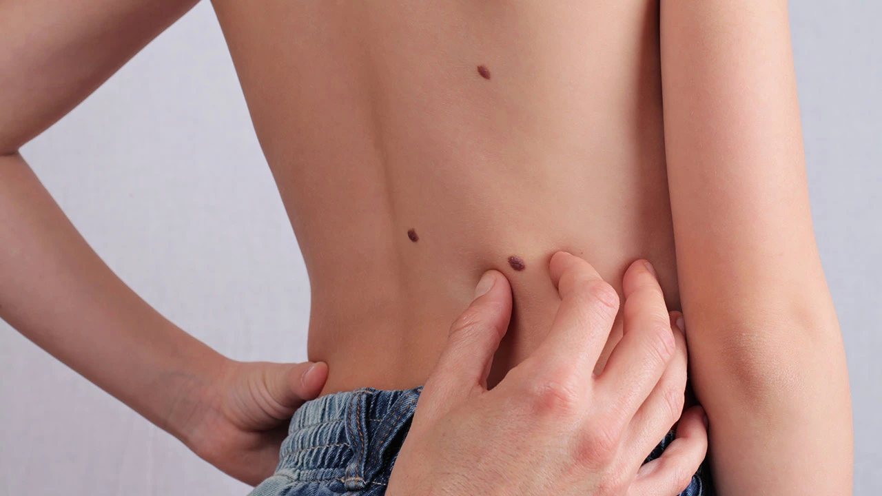
Which types of moles can be removed from the skin?
All types of moles can be removed from the skin. The important factors to consider are the location, size, and type of the mole. Meticulous planning and management of treatment are required for moles located near important structures (such as the eye), moles larger than 5-10mm, and moles with a higher likelihood of malignancy (cancerous). Particularly for moles suspected to be malignant, exhibiting the following characteristics:
- Asymmetry
- Irregular borders
- Multiple colors (shades of light, dark, red, brown, etc.)
- Diameter larger than 5mm
- Evolution, showing signs of growth, shape change, or bleeding
- For such moles that exhibit these features, it is essential to send them for pathology examination. Additional treatments may be required based on the pathology results.
The most commonly removed benign moles from the skin include skin tags, dermal nevi, melanocytic nevi, spitz nevi, solar lentigo, syringoma, milia, and seborrheic keratosis.
The most commonly removed benign lesions from beneath the skin include lipomas (fatty tumors) and sebaceous cysts (epidermoid cysts).
The most commonly removed malignant tumors from the skin include basal cell carcinoma (BCC), squamous cell carcinoma (SCC), malignant melanoma, and Merkel cell tumor.
Can moles grow back after they are removed from the skin?
In more than 90% of cases, moles do not grow back after they are removed from the skin. However, there is no guarantee that new moles won’t appear in the same area or elsewhere on the body. Although it is not commonly observed in practical applications, moles can develop anywhere on the body, including areas where moles were previously removed.
What can be expected after mole removal Turkey from the skin? What types of results can be achieved?
The skin can be performed for two reasons: cosmetic purposes or medical reasons. For cosmetic purposes, the aim is to get rid of the unsightly and noticeable appearance of the mole. In some cases, even if there is minimal scarring, the outcome can result in a much more aesthetically pleasing appearance compared to the mole itself.
Especially when there are moles that are frequently touched, scratched, or cause irritation for oneself or children, their removal is recommended for both quality of life and to reduce the chances of these constantly irritated lesions turning into malignant lesions in the future. Medically, moles suspected of being malignant must be removed and sent for pathology examination. Early diagnosis is crucial, particularly for skin cancers like malignant melanoma, as it can save lives.
Which specialist should be consulted for mole removal Turkey?
Mole removal Turkey procedures are performed by Plastic, Reconstructive, and Aesthetic Surgeons in most public institutions and private hospitals. Dermatologists who have specialized in this field and other surgical specialists also intervene, especially for moles located in visually significant areas like the face. Procedures performed by unqualified individuals (such as applying black salve, etc.) can result in excessive scarring and delay the diagnosis of malignant diseases.
Mole Removal Turkey Cost 2024
The prices for mole removal Turkey from the skin are determined according to the Istanbul Medical Chamber’s 2024 Mole Removal Turkey cost list. The fees vary based on the number of procedures to be performed, the type of anesthesia, pathology expenses, the size and popularity of the institutions, and the patient volume, ranging from 1 to 10 times the listed prices. For 2024 mole removal prices at our clinic, please contact us.
How should skin care be done after mole removal Turkey?
Wound care should be performed until the wound heals, and after the wound has healed and the stitches are removed, normal skincare can be resumed in addition to scar-reducing creams.
What steps should be followed for mole removal from the skin?
For mole removal from the skin, you can visit the nearest Dermatology or Plastic, Reconstructive, and Aesthetic Surgery specialist for an examination. (Best doctors for mole removal Turkey)
When can normal activities be resumed after mole removal?
The recovery period after mole removal Turkey depends on the treated area and the individual’s overall health. After mole removal Turkey from the facial area, it is recommended to avoid activities that may increase blood pressure or strain the stitches for about a week. This may be longer for lower extremities (thighs, legs, feet). Once your stitches are removed, you can return to your normal activities. If your mole was removed using non-stitch ablative methods, you can return to your normal activities after 4-5 days, provided you take proper care of the wound.
Of course, specific questions and concerns may arise based on individual circumstances and preferences. It is important to consult a qualified dermatologist or plastic surgeon to address your personal concerns and obtain accurate information tailored to your situation.